Worldwide Presence

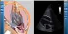
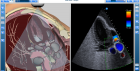

VIMEDIX transforms learning for echocardiography
VIMEDIX is a ground-breaking echocardiography training platform that offers on-demand learning without risk to live patients. Using computer-generated echo simulation, VIMEDIX teaches learners how to perform cardiac echo imaging procedures and interpret the scanned ultrasound images.
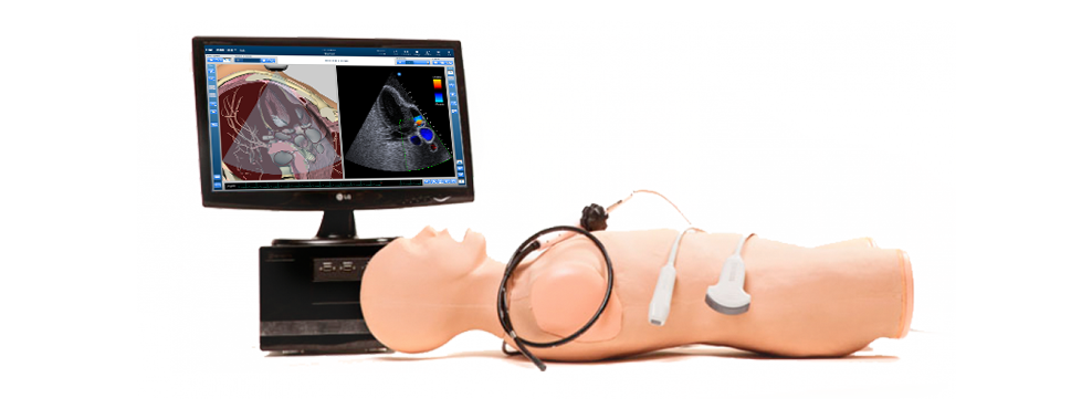
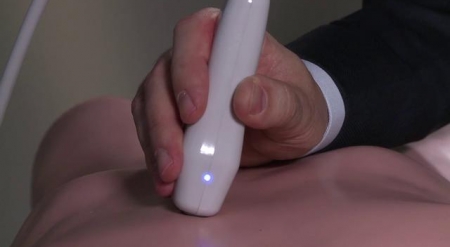
With a manikin, mock ultrasound transducer and graphic representation of both the patient anatomy and the ultrasound scan in real-time, VIMEDIX offers an immersive ultrasound simulation learning environment that shortens the path to competency. The VIMEDIX manikin’s realistic tactile features, including a depressible abdomen, ribs and sternum allow the student to practice fine motor skills associated with performing cardiac ultrasound exams.

Real-time echo simulation with augmented reality
As the learner scans the mannequin, the VIMEDIX high-resolution monitor displays a realistic simulated ultrasound image ultrasound beside a real-time 3D graphic representation of the anatomy, including surrounding structures and organs. The 3D anatomy feature can be toggled on or off, or it can be customized to adapt to the learner’s level of knowledge and ultrasound imaging skill. This augmented reality feature improves understanding of how the heartworks and speeds the path to proficiency in obtaining ultrasound images. As a learner progresses, the instructor may turn off the 360-degree anatomical image and transform the echo simulator into an ultrasound imaging system that more closely resembles what a resident would find in a clinical setting.
A realistic ultrasound simulator that can be tailored to each learner
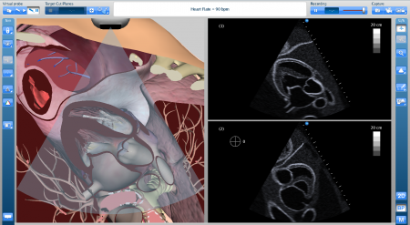
Beauty meets functionality in the newest release of the VIMEDIX ultrasound simulator user interface. Now you can customize your screen based on your user preferences and learning objectives. Beginners may view only the 3D anatomy within a semi-transparent ultrasound beam, while advanced practitioners may choose a full screen imaging mode and a more challenging ultrasound display. The new biplane imaging mode, which is used primarily for viewing the heart, displays images captured from two ultrasound beams simultaneously. Smart Metrics capture more learner movements and can display several captured target cut planes in a series. Instructors can view a side-by-side comparison of the target vs. captured images for learner assessment at-a-glance.


On-demand learning: Scan when you want, where you want without risk to live patients
Users to can choose from more than 50 normal or pathology cases and a range of body types as they practice real-time transthoracic echocardiography (TTE) and the transesophageal echo (TEE) exam with a variety of patient cases. By simply selecting a patient, an instructor can train a resident or echocardiographer how to scan a prosthetic aortic valve, assess a patient with pulmonary hypertension or perform a Focused Assessment with Sonography for Trauma (FAST) exam. With cases available on-demand, learners can gain in-depth experience and hours of psychomotor skill practice associated with ultrasound before they touch a real patient.
More than 50 Cases and Pathologies
VIMEDIX is the most comprehensive and versatile ultrasound simulator today, with more than 50 pathologies for cardiac and abdominal ultrasound assessment, including myocardial infarction, pleural effusion, tamponade, aortic aneurysm, Morrison’s Pouch, pancreatitis, renal cyst, cirrhosis and more. Educators can continually access the growing library of new cases online.


Ultrasound scanning with color Doppler imaging on more than 22 cardiac pathologies
The newest release of VIMEDIX allows users to perform color Doppler imaging on a normal heart and on 22 cardiac pathologies.
Color Doppler-enabled pathologies:
- Normal Heart
- Dilated Cardiomyopathy – Severe Biventricular Systolic Dysfunction
- Hyperdynamic Left Ventricular Systolic Function
- Recent Anterior Myocardial Infarction with Pericardial Effusion
- Dilated Cardiomyopathy – Very Severe Left Ventricular Systolic Dysfunction in a COPD Patient
- Dilated Cardiomyopathy – Mild Left Ventricular Systolic Dysfunction
- Anterior Myocardial Infarction in a COPD Patient
- Biologic Prosthetic Valve in Aortic Position
- Dilated Cardiomyopathy – Very Severe Left Ventricular Systolic Dysfunction
- Pulmonary Hypertension in a COPD Patient
- Pulmonary Hypertension
- Bicuspid Aortic Valve
- Atrial Septal Defect - small
- Dilated Cardiomyopathy – Moderate Biventricular Systolic Dysfunction
- Coronary Artery Disease – Wall Motion Abnormalities in the 3 Coronary Territories
- Left Pleural Effusion
- Normal Heart in a COPD Patient
- Tamponade
- Left Ventricular Apical Aneurysm with Thrombus
- Fine Ventricular Fibrillation
- Dilated Cardiomyopathy - Mild Left Ventricular Systolic Dysfunction in a COPD Patient
- Acute Lateral Myocardial Infarction in a COPD Patient
- Acute Anterior Myocardial Infarction
Scalable Ultrasound Simulation System for Cardiology or for Multiple Specialties
The VIMEDIX ultrasound simulation standard system includes a mannequin, mock transducer, monitor and transthoracic (TTE) and FOCUS exams. Instructors may add the transesophageal (TEE) exam and probe, the FAST module and probe and abdominal ultrasound simulation modules. VIMEDIX is used in resident programs and teaching hospitals for training in cardiovascular ultrasound, critical care and emergency medicine ultrasound and anesthesia ultrasound. The new VIMEDIX Ob/Gyn Ultrasound simulator can be purchased as an add-on system for the practice of obstetrical ultrasound.


Onsite Ultrasound Training
CAE Healthcare offers a variety of ultrasound training courses designed to teach basic principles of the sonographic exam being used as an extension to the physical examination. Courses are addressed to physicians with or without prior ultrasound experience, including intensivists, ER physicians, anesthesiologists, surgeons, cardiologists, nurse practitioners, etc. All courses are provided by highly trained critical care physicians and certified sonographers. Courses can be given directly at your medical facility and are recognized for CME credits by the Royal College of Physicians, the American Medical Associations and the Universite de Montreal.